Research Center
Leadership
Elizabeth Laugeson, PsyD
Director, UCLA Tarjan Center
Contact Info
UCLA Tarjan Center Website
760 Westwood Plaza
58-228 Semel
Los Angeles, CA
tarjancenter@mednet.ucla.edu
About
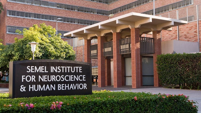
The UCLA Tarjan Center is one of 68 federally designated University Centers for Excellence in Developmental Disabilities (UCEDD) authorized under the Developmental Disabilities Assistance and Bill of Rights Act. Since the 1960s, the Tarjan Center has been a cornerstone of service, innovation, and advocacy in the disability community. Our strategic location within the Semel Institute facilitates collaboration and knowledge-sharing with cutting-edge clinical and research programs. Many faculty members hold positions within the David Geffen School of Medicine, Department of Psychiatry and Biobehavioral Sciences.
The Tarjan Center serves as a bridge between the resources of the university; local, state and international organizations; agencies; policy makers; people with disabilities; and their families. We provide training, technical assistance, community service, research, and information dissemination. All of our efforts are aimed at advancing health and wellness (both mental and physical), supporting postsecondary education, and improving employment outcomes for individuals with developmental disabilities.